Skin lesion removal by consultant plastic surgeons and laser specialists
Skin lesions such as skin tags, moles, cysts and lipomas are often removed for aesthetic reasons. But other lesions may be of concern and need assessing by a skin cancer specialist. All our consultant plastic surgeons are skin cancer experts and can assess your lesion using a dermatoscope, which is a visual aid used when examining and diagnosing lesions such as melanoma or other suspicious growths. We also have pathology diagnostic services available so your lesion can be tested for anything of concern, if required.
Description
Skin lesion removal can be performed as a non-surgical or minor surgical procedure at our aesthetic facility in Newcastle. If requested, or advised, the lesion can be sent for histological analysis to ensure it is entirely safe. If necessary, referral to the NHS can be made by our clinical team.
Cost
Skin Lesion Removal
See & Treat Clinics – Surgical
- £500 for one lesion
- £650 for up to three
Skin Consultation Only: £100–£150
Frequently Asked Questions
Making the decision to undergo surgical procedures can be a difficult one, and there are many questions you may have. Please read the frequently asked questions below which may answer any queries or ease any concerns you may have.
What is a skin lesion?
A skin lesion may be a mole (nevi), skin tag, wart, cyst, actinic (solar) keratosis, seborrheic keratosis, lipoma. More suspicious lesions include basal cell carcinoma (BCC), squamous cell carcinoma (SCC) and melanoma.
Does it hurt?
Direct excision, shave excision and laser removal are all carried out under local anaesthetic. This is administered through a tiny needle to minimise discomfort, once the anaesthetic is working, you will not feel any pain or discomfort during the procedure. Cryotherapy does not require local anaesthetic as any very minor discomfort is short lived.
Can I drive home afterwards?
Normally, you should be able to drive home after a lesion removal procedure with local anaesthetic. Since this only numbs the area being treated and doesn’t affect your overall mental state or motor skills, you should feel fine to drive afterwards.
If you were having multiple lesions removed, your surgeon may advise you to organise transport after your procedure.
Is the lesion assessed by a pathologist?
Once you have been assessed by one of our consultant plastic surgeons, they will discuss with you if they feel pathology is advisable. If so, the lesion is sent for analysis. Once we receive your result, your surgeon will convey the report to you.
What are the risk factors of lesion removal?
The risks of lesion removal include infection, bleeding, pain, scarring at the site of the lesion, possible thick keloid scarring, incomplete excision or removal, reoccurrence, contour defects and hypo or hyperpigmentation.
Understanding Different Types of Skin Lesions and Their Removal Methods
Skin lesions are abnormal changes in the skin’s texture or colour. They can be caused by infections, allergic reactions, or underlying medical conditions. While some are harmless, others may require medical attention. Here, we’ll explore different types of skin lesions and how they can be removed.
Types of Skin Lesions
1. Moles (Nevi)
Moles are common pigmented growths on the skin. They are usually harmless but can sometimes develop into melanoma, a type of skin cancer. Monitoring changes in size, colour, or shape is crucial.
Removal Methods:
• Surgical Excision: The mole is cut out, and stitches are used to close the wound.
• Shave Removal: A scalpel is used to shave off the mole at skin level.
• Laser Removal: Light energy breaks down the tissue and pigment in the mole, gradually reducing the size until flat.
2. Skin Tags
Skin tags are small, benign growths commonly found on the neck, underarms, and eyelids. They are painless but may be removed for cosmetic reasons or discomfort.
Removal Methods:
• Cryotherapy: Freezing the skin tag with liquid nitrogen.
• Cauterization: Burning off the skin tag using an electric current.
• Excision: Cutting off the skin tag with surgical scissors.
• Laser: Controlled heat using an ablative laser will remove the skin tag.
3. Warts
Caused by the human papillomavirus (HPV), warts appear as rough, raised bumps on the skin. They are contagious and can spread through direct contact.
Removal Methods:
• Cryotherapy: Freezing the wart off with liquid nitrogen.
• Salicylic Acid Treatments: Over-the-counter treatments that gradually break down the wart.
• Laser Therapy: Using laser energy to destroy wart tissue.
4. Cysts
Cysts are fluid-filled sacs that form beneath the skin. They can become infected or inflamed, requiring medical intervention.
Removal Methods:
• Drainage: The cyst is punctured, and fluid is drained.
• Surgical Removal: The cyst and its sac are completely removed to prevent recurrence.
• Steroid Injections: Used to reduce inflammation and shrink the cyst.
5. Actinic (Solar) Keratosis
These are rough, scaly patches caused by prolonged sun exposure. They are considered precancerous and should be treated promptly.
Removal Methods:
• Cryotherapy: Freezing the lesion to destroy abnormal cells.
• Topical Medications: Prescription creams that remove damaged skin cells.
• Laser: Controlled heat is used to flatten and lighten the lesion.
• Photodynamic Therapy (PDT): Light therapy combined with a photosensitizing agent.
6. Seborrheic Keratosis
These noncancerous skin growths appear as warty, scaly patches that can be brown, black, or tan.
Removal Methods:
• Cryotherapy
• Curettage: Scraping off the lesion.
• Laser: Controlled heat is used to flatten and lighten the lesion.
7. Lipomas
Lipomas are soft, fatty lumps that grow under the skin. They are usually harmless but can be removed if they become painful or bothersome.
Removal Methods:
• Surgical Excision
Skin Cancers
1.Melanoma
A melanoma is a type of skin cancer that develops in the cells called melanocytes, which are responsible for producing the pigment melanin that gives skin its colour. It typically begins in a mole or pigmented area on the skin but can spread to other parts of the body if not detected and treated early.
Melanomas are often characterised by changes in the appearance of a mole, such as:
- Asymmetry: The shape of the mole is uneven.
- Borders: The edges may be irregular, jagged, or blurred.
- Colour: The colour may vary, with shades of brown, black, or sometimes pink, red, or blue.
- Diameter: It may grow larger than the size of a pencil eraser (about 6mm).
- Evolving: It changes in size, shape, or colour over time.
Melanomas are less common than other types of skin cancer but are more dangerous, as they can spread quickly if not treated early. It’s important to regularly check your skin for any changes in moles. Our plastic surgeons are skin cancer specialists who can assess your lesion if you notice anything unusual.
Removal Method:
- Surgical removal: Via direct excision
2.Basal cell carcinoma
A basal cell carcinoma (BCC) is the most common type of skin cancer. It develops in the basal cells, which are located in the deepest layer of the epidermis (the outermost layer of the skin). BCCs are usually slow-growing and tend to stay confined to the area where they originated, making them less likely to spread to other parts of the body compared to other types of skin cancer.
Basal cell carcinoma is often linked to sun exposure, especially in people with fair skin, and it is more common in older adults. However, it can occur in people of all skin types.
Common signs of BCC include:
- A pearly or waxy bump: Often on sun-exposed areas such as the face, ears, neck, scalp, shoulders, and back.
- A flat, flesh-coloured or brown scar-like lesion.
- Bleeding or crusting: The growth may bleed or form a scab, especially if irritated or scratched.
- A raised, red, or irritated area that may have visible blood vessels.
Although basal cell carcinomas are generally not life-threatening, they can cause significant damage to surrounding tissues if left untreated. Early detection and treatment are important to prevent complications.
Removal Methods:
- Surgical removal: The most common treatment, often via direct excision.
- Cryotherapy: Freezing the cancer cells with liquid nitrogen.
- Topical treatments
3.Squamous Cell Carcinoma (SCC)
Squamous cell carcinoma (SCC) is a type of skin cancer that arises from the squamous cells, which are flat cells found in the outer layer of the skin (epidermis). It is the second most common type of skin cancer after basal cell carcinoma.
Causes and Risk Factors:
- UV exposure: Prolonged exposure to sunlight or tanning beds is the most significant risk factor.
- Fair skin: People with lighter skin are more susceptible.
- Age: More common in older adults.
- Weakened immune system: Conditions like HIV or immunosuppressive medications increase risk.
- Previous skin damage: Scars, burns, or chronic wounds can predispose to SCC.
- HPV infection: Certain strains of the human papillomavirus (HPV) are linked to SCC, particularly in mucosal areas.
Symptoms:
- A scaly, red, rough, or crusty patch on the skin.
- A sore that does not heal or keeps returning.
- A raised growth with a central depression.
- A wart-like growth.
- Itching, tenderness, or bleeding in the affected area.
Removal Methods:
- Surgical removal: The most common treatment, often via direct excision.
- Cryotherapy: Freezing the cancer cells with liquid nitrogen.
- Radiotherapy: Used for cases where surgery is not an option.
- Topical treatments
When to Seek Medical Advice
Seek medical advice if you notice:
• Rapid growth or changes in a lesion
• Irregular borders or multiple colours in the lesion
• Pain, bleeding, or itching
• Persistent lesions that do not heal
All our surgeons at LASE Cosmetic are skin cancer specialists. If you are concerned or simply want reassurance, we are here to help. You can either send in a selfie surgery photo from our website or call the clinic to organise an assessment.
You may need a face to face skin consultation, but, for simple moles and lesions that do not require pathology, it may be most appropriate to have the lesion removed by laser. This of course, rules out any option for pathology but it is a simple and easy way to remove lesions, particularly for aesthetic purposes.
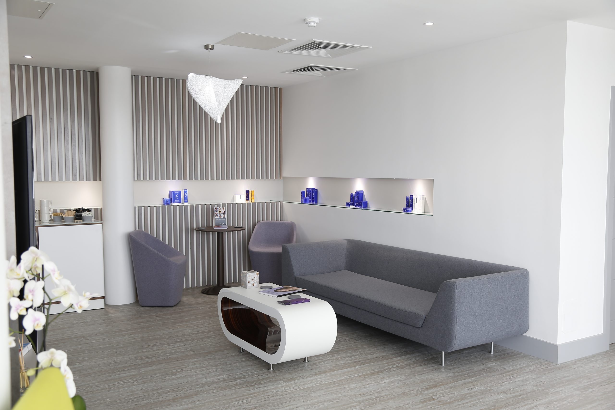
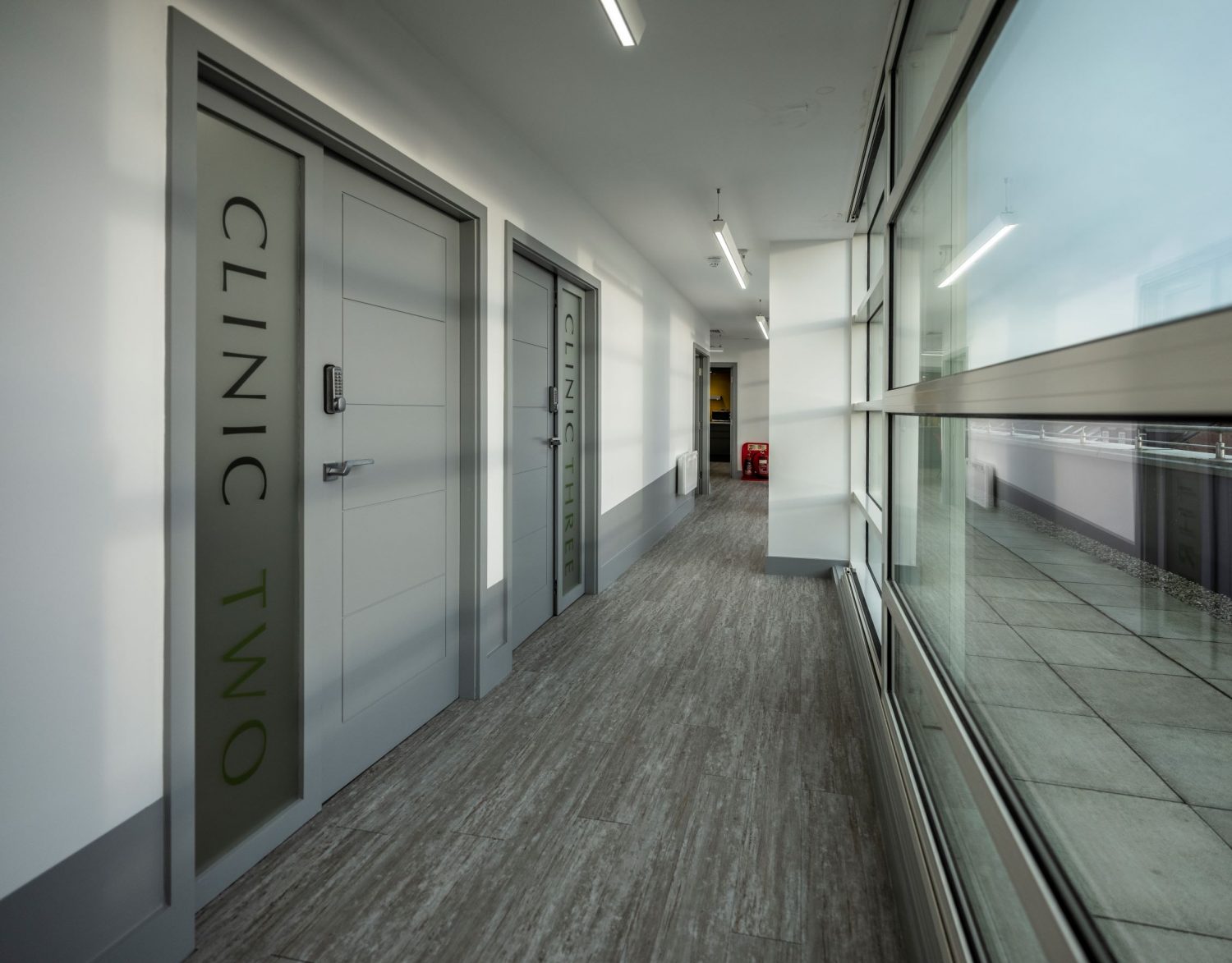
Our Clinic


Get In Touch
Follow Us